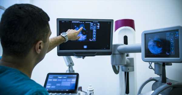
Combining AI-powered image analysis with image-based risk scoring has demonstrated the ability to predict long-term breast cancer risk, according to a new study from the Mayo Clinic and the University of California, San Francisco.
WHY IT MATTERS
In the report, published in the Journal of Clinical Oncology, researchers used Screenpoint Medical’s Transpara Exam Score with Volpara’s TruDensity software to study how the combination can assist radiologists in predicting the long-term risk of advanced and interval breast cancers.
Mayo Clinic and UCSF pulled two-dimensional full-field digital mammograms performed 2-5.5 years before cancer diagnosis from 2,412 women with invasive breast cancer and 4,995 controls from two US mammography cohorts.
They found that using AI-powered image analysis with an image-based risk tool that categorizes exams would enable radiologists to quantify breast tissue with precision, as the image software reduces reader variability in assessing breast density.
The TruDensity AI algorithm uses a combination of X-ray physics and machine learning to generate an accurate volumetric measure of breast composition to eliminate variability that can arise from human interpretation, according to the announcement about the new U.S. study.
“Breast density is a critical factor in assessing breast cancer risk, and an objective, volumetric measurement of density is pivotal,” said Dr. Ralph Highnam, Volpara Health’s chief science and innovation officer, in a statement.
“Through the power of AI, we can uncover valuable insights that help clinicians identify individuals at risk for cancer and tailor personalized screening and prevention strategies.”
This retrospective study echoed a previous study published in March in European Radiology that merged screening outcomes from women attending BreastScreen Norway with AI scores and explored consensus, recall and cancer detection for different theoretical scenarios of AI and the radiologists in screen-reading.
The Norwegian study tested the approach – using Transpara’s exam scores and TruDensity’s image analysis – to evaluate 949 screen-detected breast cancers, 305 interval cancers and 13,646 negative examinations performed between 2010 and 2018.
“We assessed Breast Imaging Reporting and Data System density, an AI malignancy score (1-10), and volumetric density measures,” the American researchers said in their abstract.
“We used conditional logistic regression to estimate odds ratios, 95% CIs, adjusted for age and BMI and C-statistics to describe the association of AI score with invasive cancer and its contribution to models with breast density measures,” they said.
While the AI score improved the prediction of all cancer types in models with density measures and discrimination improved for advanced cancer, the approach did not reach statistical significance for interval cancer, or breast cancer detected between mammography screenings.
The Norway researchers noted that 20–30% of the screen-detected and interval cancers are classified as missed in retrospective informed review studies, and cited the double reading protocol and the shortage of European radiologists as driving the need for AI-driven diagnostic tools.
“AI systems have been proposed as a tool to support or even replace radiologists in the reading process,” they wrote in the full report. They were able to compare the performance of AI against the radiologists’ double reading.
“The accuracy of the AI system was comparable to one reader in an independent double-reading setting,” they said.
THE LARGER TREND
For several years, the battle against breast cancer has been complicated by reading mammograms in women with high levels of breast tissue density. The drive is to advance early detection and get women into care faster – and suppress rising breast cancer mortality rates worldwide.
Breast cancer is not the only area where artificial intelligence can help radiologists, and many fear AI will replace doctors.
While the immediate and long-term benefits of AI can advance a diagnosis and then drive care management decisions, providers should rest assured, according to Dr. Michael Donovan, cofounder and chief medical officer at PreciseDx, a health IT company that uses AI to personalize medicine.
“A key point is that, through these advantages and efficiencies of care provided by AI to the physician, the ultimate arbiter, from a diagnostic, ethical and medical legal perspective, is the physician,” he told Healthcare IT News in January.
“For example, AI alone can’t make the final diagnosis on a radiographic scan or provide a definitive diagnosis on a patient’s image of their needle-biopsy specimen.”
ON THE RECORD
“While we have known for decades that density and breast cancer risk are correlated, recent research has really pushed forward our ability to better understand the effects of density combined with image-based risk to drive personalized medicine for women,” Dr. Nico Karssemeijer, chief scientific officer at Screenpoint Medical and professor at Radboud University, said in the statement.
Andrea Fox is senior editor of Healthcare IT News.
Email: [email protected]
Healthcare IT News is a HIMSS Media publication.
Source: Read Full Article
