
Outer retinal band (ORB) integrity and outer retinal thickness as they appear on OCT (Optical Coherence Tomography) have been studied extensively due to their diagnostic value. They are predictive of visual outcome in many retinal diseases like age-related macular degeneration (AMD), diabetic retinopathy, retinal detachment, or retinal degeneration. Despite the common use of various animal species in preclinical ophthalmic research involving OCT, no consensus exists regarding the nomenclature of ORBs as imaged by OCT in different species. On the contrary, the identification/nomenclature of ORB and the definition of the retinal/choroidal junction on OCT in various species in the scientific literature are contradictory.
A study now published in the ARVO journal Investigative Ophthalmology & Visual Science shows: The choriocapillaris is an easy and valid marker for identification of the outer retinal margin in four common experimental animal species (minipig, rabbit, rat, mouse). Peter Maloca, Group Leader Ophthalmic Imaging at IOB, is shared first author of the paper.
Sounds boring? Irrelevant?
This is both exciting and important for translational ophthalmic research. Proper and consistent outer retinal margin and ORB identification is essential for research result reproducibility and translation into new therapies for blinding diseases. Correct identification of the outer retinal margin is vital for repeatable retinal thickness measurements and proper identification of ORB on OCT images. Moreover, it is essential for reproducibility of research results, translation of animal data to humans and correlation of OCT and histology data. It matters because animal models are still indispensable in translational ophthalmic research to date, despite the availability of human retinal organoids in a petri-dish.
The study at a glance
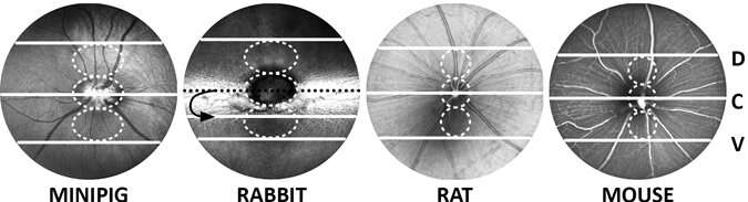
The purpose of this study was to assess with spectral-domain optical coherence tomography (OCT) the interspecies variation of outer retinal morphology and identification of the choriocapillaris in four research animal species. 574 B-scans from 96 subjects in four common experimental animal species (minipig, rabbit, rat, mouse) were evaluated. The percentage of OCT B-scans with an identifiable choriocapillaris band in the superior, central, and inferior retina were quantified. OCT images were evaluated by two independent readers. OCT findings were correlated with histology.
What is OCT?
Source: Read Full Article