Researchers are constantly looking for new antibodies that show effectiveness against severe acute respiratory syndrome coronavirus 2 (SARS-CoV-2), as they have multiple applications in testing, research, and therapeutics. Researchers from the Biological Defence Programme of Singapore have investigated six of these antibodies.
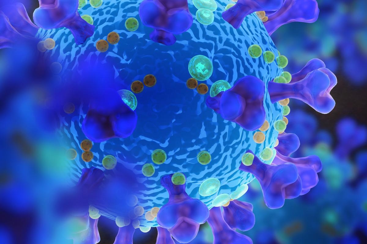
 Study: The cellular characterisation of SARS-CoV-2 spike protein in virus-infected cells using Receptor Binding Domain-binding specific human monoclonal antibodies. Image Credit: Dotted Yeti/Shutterstock
A preprint version of the study is available on the bioRxiv* server while the article undergoes peer review.
The study
The researchers used three strains of SARS-CoV-2 isolated from Singapore in the early days of the pandemic. These are SARS-CoV-2/1302, SARS-CoV-2/0536, and SARS-CoV-2/0334, referred to in the future as 1302, 0536, and 0334, respectively.
Following isolation, the complete spike protein sequence for each strain was analyzed. The data revealed that all three sequences had a high level of homology compared to the spike protein sequence of the WIV04 strain, the wild-type strain found in Wuhan, China. These isolates showed more differences in the primary amino acid sequences with each subsequent passage. 0334 and 0563 were identical, and both showed a five amino acid deletion just upstream of the furin cleavage site. This was not observed in 1302, which instead showed a single amino acid change within the cleavage site. The remaining spike protein sequences in all strains were identical to wild-type – including the sequences of the receptor-binding domain (RBD).
The researchers chose to move forward with analyzing 1302 and 0334. They selected five antibodies, PD4, PD5, PD7, SC23, and SC29, and attempted to detect the binding of the antibodies to the spike protein using Western blotting. Unfortunately, this failed, and the scientists moved on to using ELISA assays to confirm recognition of the spike protein. PD5, PD7, and SC23 all showed the ability to bind the RBD.
The researchers predict that the Western blot processing altered the conformation of the spike protein, preventing antibody binding. They argue that this is evidence towards the idea that RBD-binding antibodies require specific spike protein conformations. This view is supported by previous studies showing that when the proportions of the conformation of the spike protein are altered, the intensity of the immune response is also altered.
The SC23 showed the lowest binding affinity for the RBD, potentially indicating that it can only recognize a distinct sequence within the RBD or requires sequences outside to facilitate binding. Both PD4 and SC29 showed no RBD binding, suggesting their recognition sequences lie elsewhere. ELISA-based competition assays showed that the PD4 and SC29 antibodies could not bind to the spike protein simultaneously, indicating that the binding sequences are very similar or overlapping. None of the other antibodies interfere with the binding of these two antibodies, but PD5, PD7, and SC23 all showed mutually exclusive binding – once again suggesting that they share similar binding sequences. Only PD5, PD7, and SC23 showed any neutralization activity when virus neutralization assays were performed.
Following this, the researchers examined the immune-reactivity of each antibody by either mock-infecting or infecting cells with 0334 and then staining them with one of the six antibodies. IF microscopy revealed that fluorescence staining was only detected in the virus-infected cells, showing the antibodies could successfully recognize the spike protein.
SEM analysis indicated that SARS-CoV-2 viral particles were present in large numbers on the surface of Vero E6 cells, indicating that a high level of virus infectivity is cell-associated. On average, more than 200 viral particles were detected per cell, although there were consistently lower numbers on the 1302 infected cells than the 0334 cells.
The scientists theorize that the 1302 virus is assembled much slower than the 0334 cells and infected monolayers of Vero E6 cells to investigate further. They stained the cells and imaged them using IF microscopy, revealing that at all moi values, both isolates showed an antibody staining pattern with clusters of stained cells, with 1302 showing consistently and significantly smaller clusters – which indicate that cell to cell transmission could be reduced with this strain as well.
The researchers also examined if furin cleavage of the spike protein was necessary to recognize the RBD by PD5. They used a furin inhibitor to prevent cleavage and examined the cells with immunoblotting cell lysates to ensure it worked. They still saw many stained cells, indicating that PD5 could still identify the RBD without cleavage.
Conclusion
The authors highlight that they have shown that three of these antibodies can recognize the RBD of the spike protein, albeit in some cases only a subpopulation of the protein that has a specific conformation. The staining patterns indicate that PD5 is characteristic of these antibodies, and it can bind to the spike protein without cleavage. This information could be valuable for researchers looking to use SARS-CoV-2 reactive antibodies for testing or therapeutics.
*Important notice
bioRxiv publishes preliminary scientific reports that are not peer-reviewed and, therefore, should not be regarded as conclusive, guide clinical practice/health-related behavior, or treated as established information
-
Chan, C. et al. (2021) "The cellular characterisation of SARS-CoV-2 spike protein in virus-infected cells using Receptor Binding Domain-binding specific human monoclonal antibodies". bioRxiv. doi: 10.1101/2021.12.06.471528. https://www.biorxiv.org/content/10.1101/2021.12.06.471528v1
Posted in: Medical Science News | Medical Research News | Disease/Infection News
Tags: Amino Acid, Antibodies, Antibody, binding affinity, Cell, Coronavirus, Coronavirus Disease COVID-19, Fluorescence, Immune Response, Kidney, Microscopy, Pandemic, Protein, Receptor, Research, Respiratory, SARS, SARS-CoV-2, Severe Acute Respiratory, Severe Acute Respiratory Syndrome, Spike Protein, Syndrome, Therapeutics, Virus, Western Blot

Written by
Sam Hancock
Sam completed his MSci in Genetics at the University of Nottingham in 2019, fuelled initially by an interest in genetic ageing. As part of his degree, he also investigated the role of rnh genes in originless replication in archaea.
Source: Read Full Article
