
Associate Professor Takeshi Kawano, Department of Electrical and Electronic Information Engineering, Toyohashi University of Technology (TUT) and the research team at the Electronics-Inspired Interdisciplinary Research Institute (EIIRIS) have developed a coaxial cable-inspired needle-electrode with the diameter of less than 10 μm, using crystal growth of semiconducting material of silicon. The microscale coaxial needle-electrode has two electrodes in the needle, enabling differential recordings to be made at very close distances, a task that had previously been difficult with conventional electrode devices. In addition, the microscale electrode reduces tissue damage compared to conventional electrodes. These advantages of the coaxial electrode enable high-quality recording of neuronal signals that could not be realized with conventional techniques, and it is expected that the coaxial needle-electrode can be used as a new way of electrophysiology in the field of neuroscience.
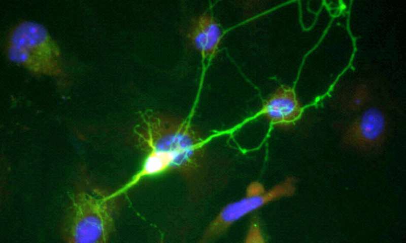
Neuronal signals can be detected by penetrating a fine electrode into the brain tissue. They are a very important technology for electrophysiological recording in the brain tissue, and by taking the advantage of high spatial resolution it is possible to gain detailed information about neuronal activity. For example, in brain-machine interface (BMI) technology—a technique that allows a patient to move their prosthetic arm or leg using signals from their brain—the technology used to implant the electrodes in the patient’s brain and record the neuronal signals with high spatial resolution is very important. It is also important to maintain a high signal-to-noise ratio when recording the signals. Electrical signals from neurons are extremely small, on the order of tens of μV (1/100,000 of 1 V), and the quality of the signal deteriorates due to noise propagating in the tissue space. This means that electrode devices must have high spatial resolution and be highly resistant to noise. Furthermore, an electrode geometry of 10 μm or less is required in order to avoid damage to the brain tissue.
To solve these challenges relating to the electrodes, the research team used the vapor-liquid-solid (VLS) growth method—a silicon growth technology—to develop a needle-like electrode device having closely positioned two electrodes in the < 10-μm-diameter needle, something that has never been achieved before. The team used an electrode device fabricated in this way to perform local-differential recording of neuronal activity with the two electrodes spaced 6 μm apart. As a result, the team achieved high-quality neuronal signal acquisition with a high signal-to-noise ratio for the first time in the world.
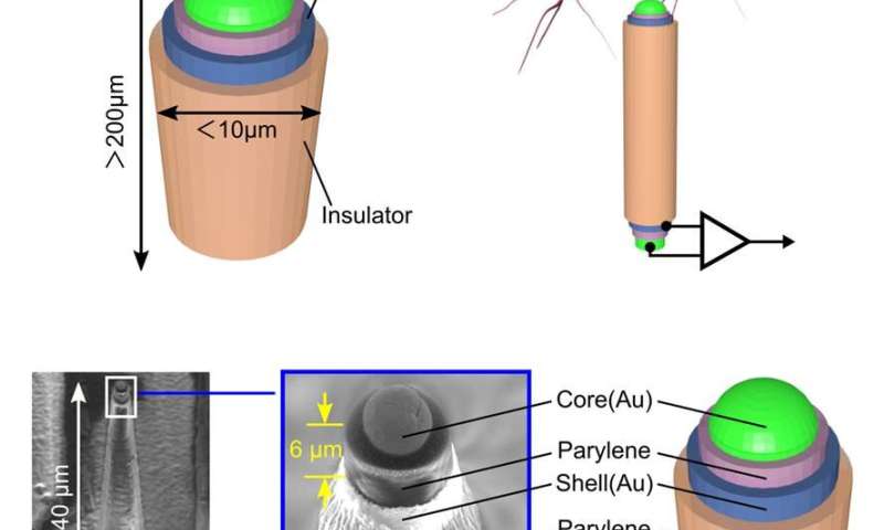
Shinnosuke Idogawa, a Ph.D. student at TUT and the lead author, commented, “In order to achieve the proposed local-differential recording, we proposed a coaxial cable-inspired microneedle-electrode. This coaxial electrode enables to dramatically reduce the electrode spacing to 6 μm compared to, for example, around 200 μm electrode spacing where conventional needle electrodes are arranged side by side. Furthermore, by performing local-differential recording with these two electrodes, we were able to reduce noise during the recording. In addition, because of the tiny needle electrode with the diameter of less than 10 μm, we are able to reduce tissue damage compared to conventional electrodes with diameters of 50 μm or more. Consequently, we have developed an electrode device that can record neuronal signals with high-quality and low invasiveness.
Source: Read Full Article