What is bioluminescence?
The luciferin-luciferase system
Bioluminescence imaging
Applications in cancer research
References
Further reading
Bioluminescence systems are widely used in cancer research, particularly in preclinical studies conducted in both in vitro and in vivo models of cancer and metastasis.

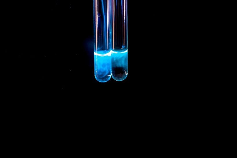
Image Credit: Emre Dikici/Shutterstock.com
What is bioluminescence?
By definition, bioluminescence describes the natural chemical reaction produced by living organisms and leads to the emission of visible light. The phenomenon of bioluminescence was first reported by Aristotle and subsequently proven true by various scientists throughout history.
These include Robert Boyle’s observations in 1667 on the importance of oxygen for bacterial bioluminescence, as well as Louise Dubois’ research on the bioluminescent beetle Pyrophorus sp. Taken together, these light-emitting proteins that are present in these organisms are referred to as “photoproteins.”
The luciferin-luciferase system
One of the most common chemical pathways used for the emission of light is the luciferin-luciferase system. Herein, luciferin acts as the substrate and light-emitting molecule, whereas the luciferase enzyme oxidizes luciferin in the presence of oxygen and/or certain cofactors such as calcium (Ca2+) or magnesium (Mg2+). The amount of light produced through this reaction is directly related to the amount of luciferin available for binding.
Notably, the bioluminescence generated by the luciferin-luciferase system can penetrate both skin and tissues. This characteristic has allowed researchers to incorporate the luciferin-luciferase system into bioluminescence imaging to assess physiological processes and diagnostics.
Bioluminescence imaging
Some of the key advantages of bioluminescence imaging include its high sensitivity, resolution, and selectivity. As a result, this non-invasive imaging modality has been widely used for studying physiological, biochemical, and pathological processes in vivo. In this regard, some notable applications of bioluminescence imaging include the labeling of cells, viruses, and bacteria, diagnosis and treatment of various diseases, assessment of protein-protein interactions, as well as studying gene expression.
Applications in cancer research
For these applications, luciferase genes cloned from various organisms will be used. Some of the most common genes used for this purpose include firefly luciferase, Renilla luciferase, Gaussian luciferase, and Vargula hilgendorfii luciferase, with firefly luciferase among the most commonly used of these genes.
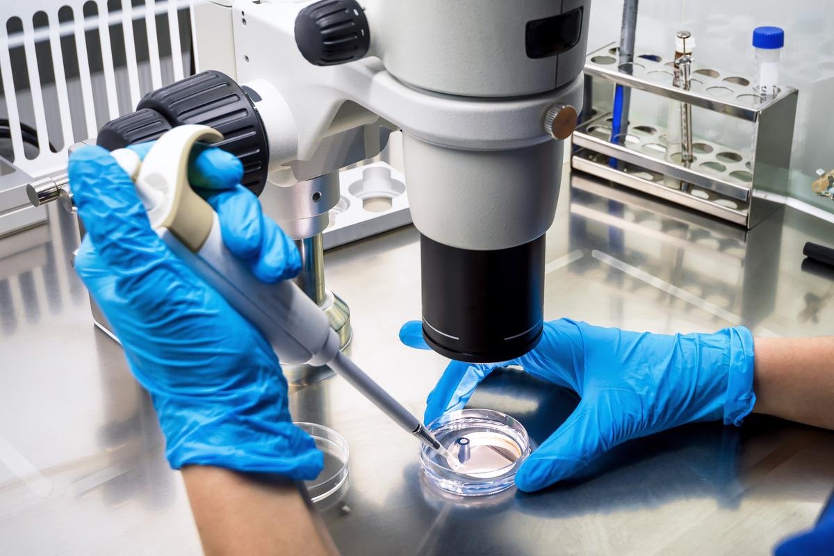
Image Credit: Viacheslav Lopatin/Shutterstock.com
Monitoring tumor growth in vivo
Many laboratories will often engineer cell lines and primary cultures to express one of these luciferase genes for cancer research stably. This is often achieved through transfection of a DNA construction of infection of the cells with lentiviral vectors.
The injection of luciferase-expressing cancer cells into various animal models is one of the most common ways in which bioluminescence is utilized in cancer research. The type of cancer being studied will often determine the injection modality selected in a given animal model.
Glioblastoma research, for example, will typically require the intracranial injection of luciferase-expressing cells, whereas a lung cancer study could instead select either a subcutaneous or intravenous injection of luciferase-expressing cancer cells.
Regardless of what injection approach is utilized, researchers can monitor the progression of any given cancer by subsequently administering luciferin to the treated animals. The luciferin will quickly bind to the luciferase genes within the cancer cells, thus leading to the chemical reaction product of bioluminescence.
The emitted light signal is then detected through in vivo imaging systems equipped with bioluminescent imaging capabilities and subsequently quantified to reflect the overall tumor size. Researchers can use this information to determine when treatments should be initiated, when the tumor burden becomes too excessive for the animal, or monitor the efficacy of various treatments over time.
Pharmacokinetics
In addition to its use for monitoring tumor growth and progression, cancer researchers have also utilized bioluminescence imaging to study the kinetics and biodistribution of various pharmaceutical agents in vivo. In a 2020 Small study, researchers developed a novel nanoformulation that consists of a luciferin probe conjugated onto the surface of the nanoparticle. When this nanoformulation enters the cytoplasm of a tumor cell, glutathione-induced cyclization occurs and subsequently leads to the prolonged release of luciferin into the cancerous cell.
Currently, many antineoplastic agents are associated with a wide range of adverse effects ranging from fatigue and hair loss to nausea and vomiting. Unfortunately, many of these reactions to cancer drugs are off-target effects; thus, there has been a considerable demand to develop more precise targeted agents to treat cancers to minimize the occurrence of toxic side effects.
To this end, the development of a sensitive approach allows researchers to monitor the targeted delivery of agents to ensure that they are reaching cells and organs of interest while also minimizing any potential off-target effects.
References
- Sharifen, S., Homaei, A., Hemmati, R., et al. (2018). The emerging use of bioluminescence in medical research. Biomedicine & Pharmacotherapy 101; 74-86. doi:10.1016/j.biopha.2018.02.065.
- Li, S., Ruan, Z., Zhang, H., & Xu, H. (2021). Recent achievements of bioluminescence imaging based on firefly luciferin-luciferase system. European Journal of Medicinal Chemistry 211. doi:10.1016/j.ejmech.2020.113111.
- Madero-Visbal, R. A., Colon, J. F., Hernandez, I. C., et al. (2010). Bioluminescence imaging correlates with tumor progression in an orthotopic mouse model of lung cancer. Surgical Oncology 21(1); 23-29. doi:10.1016/j.suronc.2010.07.008.
- Luwor, R. B., Stylli, S. S., & Kaye, A. H. (2015). Using bioluminescence imaging in glioma research. Journal of Clinical Neuroscience 22(5); 779-784. doi:10.1016/j.jocn.2014.11.001.
- Bellini, M., Riva, B., Tunelli, V., et al. (2020). Engineered Ferritin Nanoparticles for the Bioluminescence Tracking of Nanodrug Delivery in Cancer. Small. doi:10.1002/smll.202001450.
Further Reading
- All Bioluminescence Content
- Synthetic Bioluminescence: An Overview
Last Updated: Jun 1, 2022

Written by
Benedette Cuffari
After completing her Bachelor of Science in Toxicology with two minors in Spanish and Chemistry in 2016, Benedette continued her studies to complete her Master of Science in Toxicology in May of 2018.During graduate school, Benedette investigated the dermatotoxicity of mechlorethamine and bendamustine; two nitrogen mustard alkylating agents that are used in anticancer therapy.
Source: Read Full Article