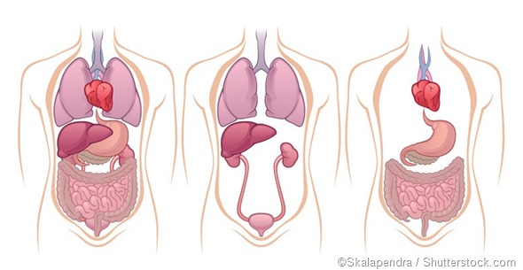
The liver and the kidneys are some of the most essential and hardworking organs in the body. They carry out numerous functions such as excretion of waste, metabolism of many substances, hormonal regulation, and proper digestion, as well as proper coagulation.

The location of the liver
The liver is the second largest organ in the body, weighing in at approximately 3 pounds. It is a deep reddish-brown in color, and is situated on the right side of the abdominal cavity, just below the right hemi-diaphragm, to the left of the stomach.
That is, it is pressing against the domed right half of the diaphragm, which separates it from the right side of the thoracic cavity. It is roughly triangular in shape, and fills almost the whole right side of the upper abdomen. Its lower border extends in front to just the level of the right ribcage margin, while at the back it extends downwards to the top of the right kidney.
The liver is covered almost completely by a fibrous capsule, and then by a peritoneal fold. The smooth glistening peritoneum encloses almost all organs within the abdomen. The peritoneum condenses in four places to form sturdy thickenings or peritoneal ligaments, which help to support the liver and keep it in place.
The surface marking of the liver is as follows:
The following three points are marked; 1.25 cm below the right nipple, 1.25 cm below the tenth rib tip and 2.5 cm below the left nipple. The line joining the first and third points marks the upper surface of the liver. The line joining the first and second points represents the lateral border of the liver. The line joining the second and third points represents the lower border.
The liver has four lobes or parts: the right and the left lobes, the caudate and the quadrate lobes.
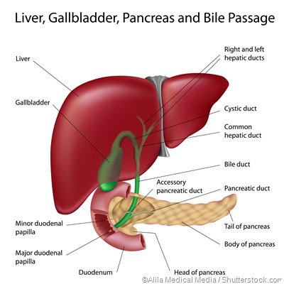
The liver cells produce bile, which is released into microscopic canals between the cells called bile canaliculi. These join as they go on to form bile ducts. Bile ducts from the different parts of the liver flow together to form the right and the left hepatic ducts, which join into the common hepatic duct.
Just below the liver, tucked into its own fossa or niche, is the gallbladder which stores the intensely bitter dark green fluid called bile. Its duct, called the cystic duct, joins with the common hepatic duct to form the common bile duct that empties bile into the duodenum as required for digestion of food.
Most of the bile that arrives via the common hepatic duct, however, goes into the gallbladder for storage till needed. Other impressions on the undersurface of the liver are made by the lowest part of the esophagus, the stomach, and the right kidney.

The location of the kidneys
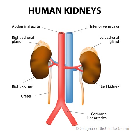
The kidneys are bean-shaped organs located in the upper retroperitoneal region of the abdomen. That is, they are located behind the smooth peritoneal lining of the upper part of the abdominal cavity, between it and the posterior body wall. Therefore, they are actually outside the peritoneal cavity.
The kidneys are situated below the diaphragm, one on either side of the spine. They are just below the rib cage. Each bears a small but crucial cap of endocrine tissue called the adrenal gland, which produce the important steroid hormones of the body.
The right kidney is approximately one centimeter lower than the left. To mark it on the body surface, the following points are used:
The upper end of the kidney is at the middle of the line joining the lower end of the sternum to the transpyloric plane – the midpoint of the line that joins the jugular notch to the upper margin of the pubic symphysis – and about 5 cm out from the center.
The lower end of the kidney is at the midpoint between the transpyloric and intertubercular planes. The latter refers to the plane that passes midway between the transpyloric plane and the upper margin of the pubic symphysis. The lower pole of the kidney is about 7 cm out from the midline.
The hilum is on the transpyloric plane, 5 cm from the midline.
On the back, the side markings are between two lines which pass 2.5 cm and 9.5 cm from the midline, respectively, while the top and bottom are marked by lines passing through the 11th thoracic and 3rd lumbar vertebrae.
The dark red kidneys handle most of the fluid waste excretion needs of the body. Each filters about 120 liters of blood plasma each and every day, cleverly separating out the waste in the form of urea and other nitrogenous substances, as well as excessive salt ions.
It keeps the body free from the toxic effects of waste build up and also maintains the even levels of salt and water in the system. The urine formed is drained into the ureter, a tube that leads downwards from the collecting part of the kidney, the renal pelvis, into the urine storage and release organ, the urinary bladder.

References
- http://www.niddk.nih.gov/health-information/health-topics/Anatomy/kidneys-how-they-work/Pages/anatomy.aspx
- http://www.ece.ncsu.edu/imaging/MedImg/SIMS/GE1_3.html
- http://www.bartleby.com/107/286.html
Further Reading
- All Kidney Content
- Renal Neoplasms
- Anatomy of the Kidney
- All Liver Content
- How do Certain Foods Affect the Liver?
Last Updated: Feb 26, 2019

Written by
Dr. Liji Thomas
Dr. Liji Thomas is an OB-GYN, who graduated from the Government Medical College, University of Calicut, Kerala, in 2001. Liji practiced as a full-time consultant in obstetrics/gynecology in a private hospital for a few years following her graduation. She has counseled hundreds of patients facing issues from pregnancy-related problems and infertility, and has been in charge of over 2,000 deliveries, striving always to achieve a normal delivery rather than operative.
Source: Read Full Article
