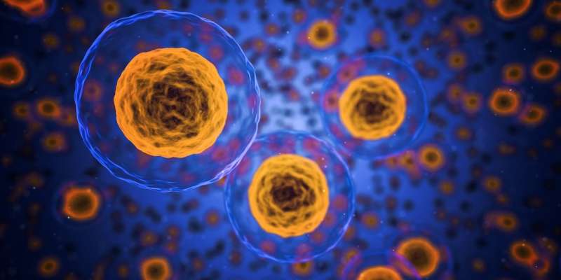
Canadian immunologists have identified a mechanism that promotes the activation of T cell receptors by altering how one of its components interacts with the cell membrane.
The finding sheds new light on the function of T cell receptors, illuminating the role of specific proteins in the cell membrane.
Up close, the T cell receptor is a cluster of proteins on the surface of T cells that bind to antigens—foreign proteins—displayed on abnormal cells, such as cancer cells or those infected with bacteria or viruses. T cell interaction with abnormal cells is a major strategy in which the immune system mounts its assault on infection, cancer, and other diseases.
As a key multi-protein complex, the T cell receptor is critical for the activation of T cells by antigens. And T cells themselves are capable of forming memories of antigens, which means upon reexposure, T cells can more rapidly respond to a familiar enemy.
Immunologists at two leading Canadian research centers, the Institut de Recherche en Immunologie et Cancérologie and the Département de Microbiologie, Infectiologie et Immunologie, Faculté de Médecine, Université de Montréal, embarked on the laboratory investigations to more fully grasp the intricate function of T cell receptors—and T cells themselves. In the end, they broke new ground, adding to the overall understanding of these cells and their storied receptors.
The new findings arrive as an increasingly bright spotlight has been trained on T cell activation, T cell binding and the role these key immune system cells play after vaccination for COVID-19. Beyond the Canadian team’s research, the subjects of T cell activation and T cell receptor biology have captured the public’s attention in the wake of the pandemic. Discussions in the popular press and social media have frequently explored whether COVID vaccination prompts memory T cell activity and a strong T cell response against SARS-CoV-2. The Montréal research has laid a new foundation of understanding for both the scientific and lay communities.
“We investigated how the electrostatic interactions that promote dynamic membrane binding of the T cell receptor-CD3 cytoplasmic domains are modulated during signaling and affect T cell activation,” Drs. Audrey Connolly and Etienne Gagnon wrote in the journal Science Signaling.
What they discovered were the critical key steps in how T cell receptor signaling is enhanced in a chain of biological events that result in the activation of T cells. The T multiprotein receptor is critical to the activation of T cells by antigens—any foreign substance that induces an immune response in the body. Although the engagement of T cell receptors by antigens activates T cell receptor signaling, T cell receptors that are not bound to antigens also can become activated and contribute to T cell activation, the team of immunologists found. T cell receptors that are not bound to antigens are called bystanders. These bystander T cell receptors serve to amplify T cell signaling.
To fully appreciate the research by the Canadian team of immunologists, it is necessary to delve into the “weeds” of structural, molecular and cellular biology to picture where these activities are physically occurring and why they are important to the immune response and other biological functions. The drama of cell signaling emanates from the surface of cells.
All cells are enclosed in the so-called plasma membrane, a thin border that surrounds every living cell. The membrane itself is made up of a lipid bilayer—two layers of phospholipids. Although this border is what maintains the integrity of cells and prevents their contents from flowing out, the plasma membrane has the consistency of olive oil. Each phospholipid bilayer is known as a “leaflet.”
The membrane as a whole is home to transmembrane proteins that are embedded in it, some protruding above the surface. Some transmembrane proteins are capable of powerful signaling activities. Channels are also in the membrane allowing the flow of ions into and out of the cell.
The Montréal immunologists were interested in a transmembrane protein from a key superfamily of transmembrane constituents—transmembrane protein 16F, or THEM16F, which is crucial to T cell activation because it aids signal amplification by the T cell receptor. The THEM16 superfamily is a calcium ion-dependent phospholipid scramblase.
Scramblases, control the distribution and composition of phospholipids within the bilayer, which has important effects on the functions of membrane proteins, by swapping phospholipids between the two leaflets.
Conolly and Gagnon found, for instance, that the scramblase TMEM16F, which redistributes phosphatidylserine from the inner to the outer leaflet of the plasma membrane, enabled CD3 cytoplasmic domains of bystander T cell receptors to disengage from the plasma membrane, leading to enhanced T cell activation. The CD3 cluster is a protein complex and T cell co-receptor involved in activating cytotoxic T cells and T helper cells.
These bystanders were in the vicinity of antigen-stimulated T cell receptors and served to amplify T cell signaling. Targeting TMEM16F therapeutically may enable modulation of T cell activation, the Montréal immunologists theorize.
In addition to playing a role in T cell activation, THEM16F also mediates the exposure of the phospholipid known as phosphatidylserine in activated platelets during blood clotting and regulates hydroxyapatite release from osteoblasts during bone mineralization. But it is the role in T cell activation that Connolly, Gagnon and colleagues sought to elucidate.
Source: Read Full Article
