
For most people, there is no scarier diagnosis than that of cancer. While treatments including chemotherapy and radiotherapy have been used since the 1940s and late 1800s, respectively, immunotherapy has more recently emerged as a viable and successful approach to cancer treatment. Indeed, evasion of the host immune system is an essential feature of tumorigenesis. Figuring out how cells do this, and disrupting it, to allow the patient’s own immune system to eliminate the cancer cells, is the basis of immunotherapy.
In a study published in August 2020 in Nature Cell Biology, a team including researchers from Tokyo Medical and Dental University (TMDU) and Harvard Medical School (HMS) have identified the regulatory mechanisms through which the PD-L1 immune check-point protein dictates the efficacy of anti-PD-L1 immunotherapy.
“We already knew that immunotherapies targeting immune-checkpoint inhibitors were somewhat successful in treating some cancer types,” says co-author Naoe Taira Nihira. “However, only a subset of patients achieved long-lasting results.”
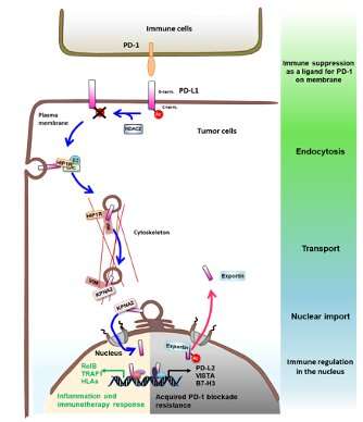
PD-L1 expression is tightly controlled, and patients with increased PD-L1 expression in tumors are likely to respond well to PD-L1 blockade; however, the reasons why increased PD-L1 expression leads to increased PD-L1 blockade sensitivity have remained unclear. The research team examined a specific kind of PD-L1 modification, called acetylation, and found that removal of this modification allows PD-L1 to enter the nucleus and interact with DNA to regulate the immune response.
Using a variety of advanced molecular, biochemical, and bioinformatics approaches, the researchers examined PD-L1 acetylation, localization, function, and interactions. They found that plasma membrane localized PD-L1 translocates to the nucleus by interacting with transport pathway components. Specifically, by introducing a series of mutations into PD-L1 and expressing different acetyltransferases, they determined that PD-L1 is acetylated by p300 at a specific residue within the cytoplasm called Lys263. Using similar approaches, and protein depletion by short-interfering RNAs, they also discovered that histone deacetylase (HDAC) specifically interacts with and deacetylates PD-L1.
Protein modifications, including acetylation, can affect protein stability, dimerization, or localization. However, when the team reduced the amount of HDAC2 protein in the cells, consequently increasing the acetylation level, there were no observable changes in protein stability or dimerization. Co-author Akira Nakanishi explains: “These results mean that the acetylation and deacetylation of PD-L1 at this residue play a critical role in its nuclear translocation.”
In the nucleus, PD-L1 regulates the expression of pro-inflammatory and immune-response-related genes, indicating that PD-L1 could function to regulate the local tumor immune environment to control its sensitivity to immune checkpoint-blockade therapy .
Source: Read Full Article