
Organ-on-a-chip technology combines engineered three-dimensional tissue within a microfluidic system to simulate the mechanics and physiology of entire organs.
XOHDY | Shutterstock
Organ-on-a-chip devices can overcome the limitations of conventional two-dimensional cell culture methodologies, allowing for the establishment of models that mimic the three-dimensional arrangement of different cell types closer to the physiological condition.
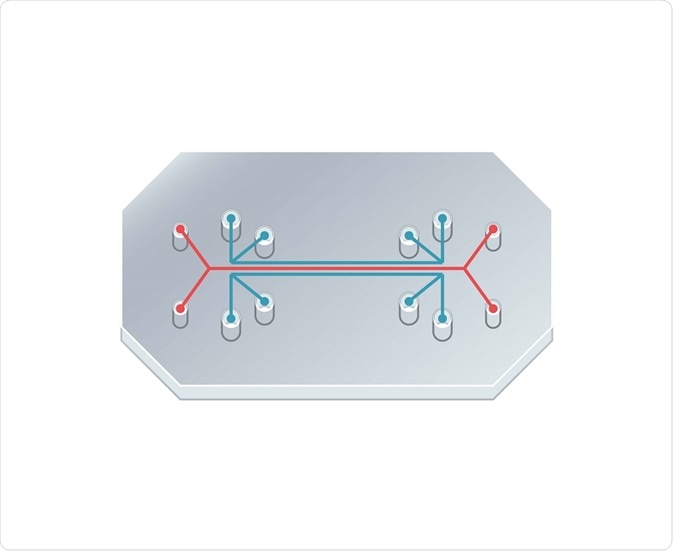
More specifically, each organ-on-a-chip is fabricated on a clear polymer containing hollow microfluidic channels lined with living human cells. The technology is still in the early stages of development but provides the possibility of drug testing without the use of animal models.
General advantages of organ-on-a-chip technology
Organ-on-a-chip technology is a significant advancement in biomedical engineering that provides multiple advantages. The formation of compartments inside the microfluidic device increases control of the microenvironment by confining cells. The laminar structure of the micro channels also allows for the formation of sub-compartments.
Such compartmentalization provides the advantage of imposing constraints to precisely control physical conditions and manipulate communication between tissue types. Organ-on-a-chip technology can also produce an optimal supply of nutrients and oxygen along with the efficient removal of catabolic metabolites.
The structure of individual organ-on-chip systems varies because the arrangement of organ specific cell types within the microfluidic device is made to resemble the basic morphology of the organ. The majority of organ-on-a-chip models utilize a porous membrane as a substrate for defining cell layers.
The membranes can, therefore, form an interface for the cellular cross talk of endothelial and epithelial cell layers. Mimicry of organ function is further enhanced by the application of mechanical forces to the device, replicating the physical microenvironment of living organs, such as the breathing motions of lungs.
Why are organs-on-a-chip used in drug development??
The high failure rate during the drug testing process is partially due to the fact that animal models are not always predictive of the human condition. Moreover, the current trial process requiring the use of animal models is expensive and time-consuming.
Considering all of the above, organ-on-a-chip technology has the potential to provide an alternative platform for drug testing. Microfluidic devices only require micro-scale volumes of cell and drug samples.
From an economic standpoint, the faster turnaround of analysis through organ-on-a-chip technology may eventually reduce the cost of drug testing. Furthermore, greater precision can be produced by the utilization of human cells.
Validation and optimization of organ-on-a-chip systems is still required before their application in drug testing. Nonetheless, there have been recent developments in the ability to connect multiple organ-on-a-chip models to analyze the effects of drug toxicity in the surrounding tissues of the target organ.
Examples of organs-on-a-chip
Microfluidic devices have been produced that simulate the functionality of organs such as the lung and heart. The lung-on-a-chip was developed to mimic the key structural, functional and mechanical properties of the human alveolar-capillary interface.
In a nutshell, the microfluidic system contains two microchannels separated by a porous membrane. Human alveolar epithelial cells and pulmonary microvascular endothelial cells are cultured on either side of the porous membrane. By compartmentalizing the channels, air can be introduced to the epithelial section to form an air-liquid interface that represents the lining of the alveolar air space.
The structure of the microfluidic device allows for the manipulation of fluid flow and the transport of nutrients. The lung-on-a-chip can also represent complex whole organ-level response, such as pulmonary inflammation, by the introduction of blood-borne immune cells to the fluid within the vascular channel.
Likewise, the heart-on-a-chip was fabricated to measure the structure-function relationships between the replicated hierarchical tissue architectures of laminar cardiac muscle. The device consists of engineered anisotropic ventricular myocardium on an elastomeric thin film.
The application of electrodes causes the contraction of myocytes (muscle cells) leading to the finding of a relationship between tissue stress and the radius of curvature produced in the muscular thin films during contraction. Recent studies indicate that the heart-on-a-chip device can be considered a valid technology for quantifying electrophysiological and contractility data from cardiac muscle tissue.
Sources:
- https://wyss.harvard.edu/media-post/introduction-to-organs-on-a-chip/
- https://ieeexplore.ieee.org/document/6678412/
- https://www.ncbi.nlm.nih.gov/pmc/articles/PMC5481808/
- http://science.sciencemag.org/content/328/5986/1662
- https://www.ncbi.nlm.nih.gov/pmc/articles/PMC4038963/
Further Reading
- All Organoids Content
- What are Organoids?
- Applications of Organoids
- Enteroids in Research
- Organoid Production Methodology
Last Updated: Dec 11, 2018

Written by
Shelley Farrar Stoakes
Shelley has a Master's degree in Human Evolution from the University of Liverpool and is currently working on her Ph.D, researching comparative primate and human skeletal anatomy. She is passionate about science communication with a particular focus on reporting the latest science news and discoveries to a broad audience. Outside of her research and science writing, Shelley enjoys reading, discovering new bands in her home city and going on long dog walks.
Source: Read Full Article